Images show ACTUAL PATIENTS. Your results may vary.
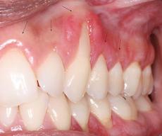
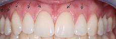
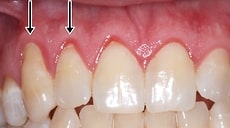
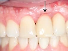
Case 62
Patient has erosive lichen planus, meaning, it is a disease whereby the top layer of the gum tissue no longer exists and is red and inflamed. 2. See arrows on pre-operative photo showing areas with gum recession. 3. 3 weeks post operative photos. Tissue from gum graft has not fully matured.
Surgeon: Dr. Alex Farnoosh
Images show ACTUAL PATIENTS. Your results may vary.
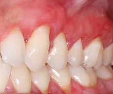
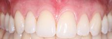
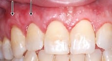
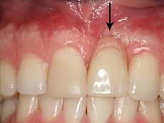
Case 61
Patient has erosive lichen planus, meaning, it is a disease whereby the top layer of the gum tissue no longer exists and is red and inflamed. 2. See arrows on pre-operative photo showing areas with gum recession. 3. 3 weeks post operative photos. Tissue from gum graft has not fully matured.
Surgeon: Dr. Alex Farnoosh
Images show ACTUAL PATIENTS. Your results may vary.
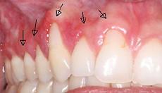
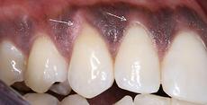
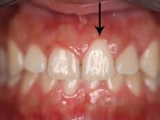
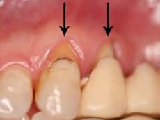
Case 60
Areas of receding gums marked with arrows were treated with AlloDerm (gum grafting material)
Surgeon: Dr. Alex Farnoosh
Images show ACTUAL PATIENTS. Your results may vary.
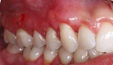
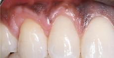
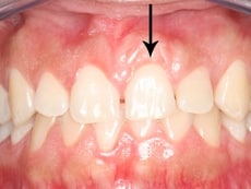
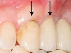
Case 59
See arrows on pre-operative photo, marking areas of receding gums treated with AlloDerm (gum grafting material)
Surgeon: Dr. Alex Farnoosh
Images show ACTUAL PATIENTS. Your results may vary.
Case 55
Gingival recession (root exposure) is caused by periodontal disease but also can be developmental, or caused by improper brushing. It is not only an aesthetic issue but also creates an area that is prone to cavities and sensitivity problems. Treatment consists of periodontal plastic surgery including a gum graft using the patient’s own tissue or utilizing Alloderm to cover the exposed root. Some common problems with previous techniques for gum grafts were scar lines, mismatched color (causing a “patchy” appearance), insufficient gum coverage, of a “bulky” appearance. Dr. Farnoosh has developed a technique that has been improved over the years to minimize the formation of the above-mentioned problems so that the graft site cannot be detected. In the “Before” image, a patient with an exposed root is seen. This patient was then treated with Dr. Farnoosh’s gum grafting technique. The “After” image was taken 4 weeks after surgery. As you can see, after proper healing, it is impossible to see that surgery has been performed. The gums look healthy and natural.
Surgeon: Dr. Alex Farnoosh
Images show ACTUAL PATIENTS. Your results may vary.
Case 56
Gingival recession (root exposure) is caused by periodontal disease but also can be developmental, or caused by improper brushing. It is not only an aesthetic issue but also creates an area that is prone to cavities and sensitivity problems. Treatment consists of periodontal plastic surgery including a gum graft using the patient’s own tissue or utilizing Alloderm to cover the exposed root. Some common problems with previous techniques for gum grafts were scar lines, mismatched color (causing a “patchy” appearance), insufficient gum coverage, of a “bulky” appearance.. Dr. Farnoosh has developed a technique that has been improved over the years to minimize the formation of the above-mentioned problems so that the graft site cannot be detected. In the “Before” image, a patient with an exposed root is seen. This patient was then treated with Dr. Farnoosh’s gum grafting technique. The “After” image was taken 4 weeks after surgery. As you can see, after proper healing, it is impossible to see that surgery has been performed. The gums look healthy and natural.
Surgeon: Dr. Alex Farnoosh
Images show ACTUAL PATIENTS. Your results may vary.
Case 57
This is a patient with dental implants and gum recession. Because this patient had dental implants, the gum recession caused a portion of the implant to become exposed, not the root. In an attempt to reduce the visibility of the metal implant and gum recession, another dentist had used stained porcelain. As you can see from the “Before” image, the stained porcelain did not achieve a good aesthetic result. Dr. Farnoosh performed gum grafting to provide significant improvement (visible in the “After” photograph).
Surgeon: Dr. Alex Farnoosh
Images show ACTUAL PATIENTS. Your results may vary.
Case 58
In these images, you see a patient with gingival recession (root exposure) on 2 teeth. To correct function and aesthetics, Dr. Farnoosh performed his special gum grafting technique to correct the root exposure without causing scar lines or discoloration. As you can see, he achieved considerable improvement.
Surgeon: Dr. Alex Farnoosh
Images show ACTUAL PATIENTS. Your results may vary.